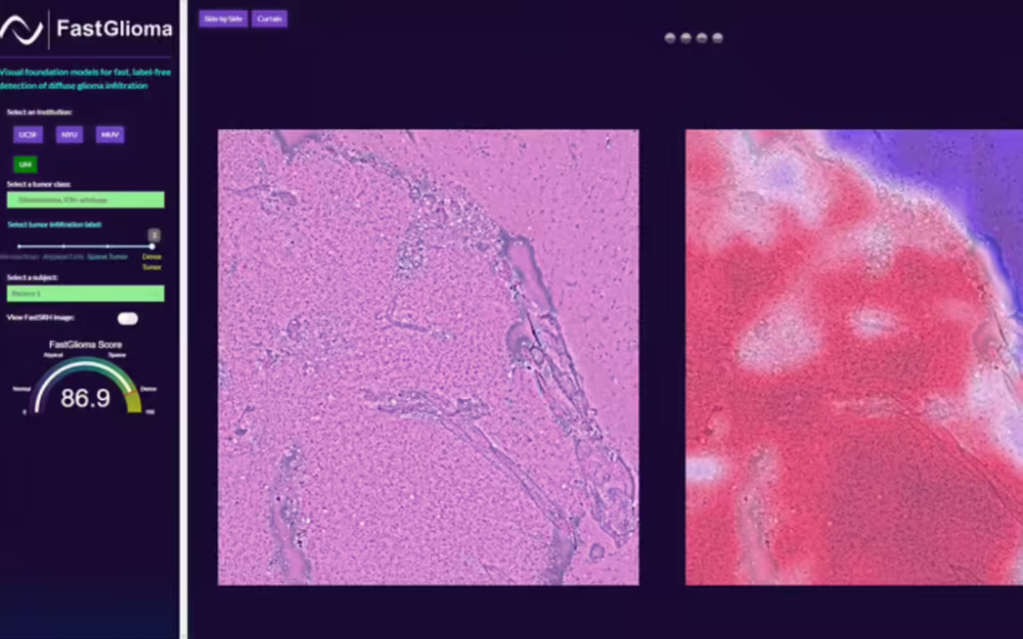
U.S. researchers have developed an artificial intelligence model called FastGlioma that can accurately identify in as little as 10 secondsbrain tumor surgeryresidual tumor tissue afterward. This breakthrough technique is expected to significantly improve the success rate of brain tumor surgery and reduce post-operative complications.

It is understood that in traditional brain tumor surgery, it is difficult for surgeons to completely remove the tumor because the line between normal brain tissue and tumor tissue is very blurred. Residual tumor tissue can lead to a range of complications, including seizures, infections, headaches, cognitive dysfunction, and motor dysfunction. While magnetic resonance imaging (MRI) and fluorescent contrast can help locate residual tumors, these methods are not always applicable to all types of tumors and may not always be feasible during surgery.
Researchers at the University of Michigan and the University of California, San Francisco, have developed a FastGlioma AI model, which was pre-trained with over 11,000 surgical samples and 4 million unique microscopic images.Detects tumor infiltration in full resolution images in less than 100 seconds with 92% accuracy. When using low-resolution images, FastGlioma can detect them in less than 10 seconds with 90% accuracy.
This technology has the potential to significantly improve patients' post-operative quality of life and reduce the need for subsequent costly corrective surgeries. In the future, the researchers plan to extend the FastGlioma model to other types of cancer, such as lung, prostate, breast and head and neck cancers, to improve the diagnosis and treatment of these cancers.